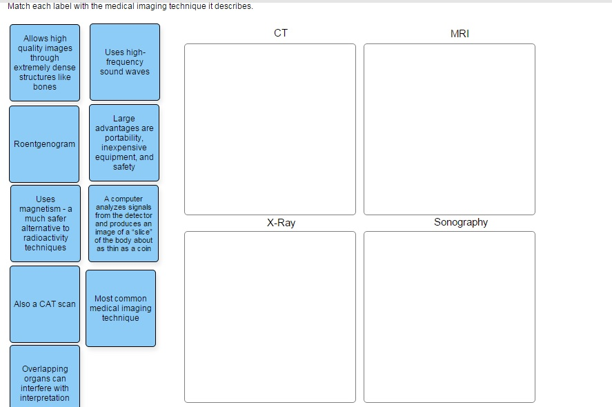
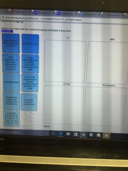
Match Each Label With the Medical Imaging Technique It Describes
These techniques include x-rays computed tomography CT scans and magnetic resonance imaging MRI. This is a way the doctor can determine if there are any abnormalities.

Match Each Label With The Medical Imaging Technique Chegg Com
Experts are tested by Chegg as specialists in their subject area.

. It has had a. Match each label with the medical imaging technique it describes. Medical imaging is a diagnostic imaging device that utilizes high-frequency sound waves to produce images of the body structure.
The grandparent of all medical imaging techniques. Terminology Basic terms of relations. Quiz 31 Medical Imaging Technologies.
With these exams doctors can see how inner organs are functioning joints are moving and much more. Use the drop-down menus to match each phrase below with the type of microscope it describes. 31 Medical Imaging Technologies 14 Terms.
Which is the most common form of medical imaging using high-energy radiation to penetrate skin and tissues but not bone. Match each label with the medical imaging technique it describes. Each kidney contains approximately 6-10 pyramids.
Is a medical imaging technique consisting of X-ray computed tomography where the X-rays are computed using cone beam technology. It uses magnetic fields and radio waves to produce these images. Medical imaging uses sound waves to look inside the body explanation BEST describes medical imaging.
Anterior is towards the front of the body Latin. In 1970 a physician and researcher named Raymond Damadian noticed that malignant cancerous. However as computer power has grown so has interest in employing advanced algorithms to facilitate our use of medical images and to enhance the information we can gain from them.
C PET scans use radiopharmaceuticals to create images of active blood flow and physiologic activity of the organ or organs being targeted. Creates a two dimensional image is used to study DNA molecules has magnification ability of up to 60000 times without losing clarity shows arrangement of atoms on surface of molecules has magnification ability of hundreds of thousands of times. Medical imaging techniques have brought about tremendous progress in the field of medicine by virtually eliminating exploratory surgeries and.
Medical imaging is the process of producing a view of the human body to diagnose or to cure the medical conditions. The brachial artery gives rise to the ulnar and radial arteries. We review their content and use your feedback to keep the quality high.
Terms in this set 24 Angiography. Match specific tooth views to specified tooth mouth windows. Match each description with medical imaging techniques.
Magnetic resonance imaging MRI is a noninvasive medical imaging technique based on a phenomenon of nuclear physics discovered in the 1930s in which matter exposed to magnetic fields and radio waves was found to emit radio signals. The techniques and procedures used to create images of the human body. These imaging tools let your doctor see inside your body to get a picture of your bones organs muscles tendons nerves and cartilage.
Medical imaging techniques such as sonography CT scans MRI scans or PET scans are one of the primary applications of body planes. A single barrage of x-rays passes through the body producing an image of interior structures on x-ray. An MRI is a diagnostic technique used to produce a detailed image of the bodies tissue and bones.
Positron Emission Tomography PET Magnetic Resonance Imaging MRI Ultrasound. Describe techniques for patient management while acquiring radiographic images including for patients with special needs. Show transcribed image text.
Medical imaging is the technique of producing visual representations of areas inside the human body to diagnose medical problems and monitor treatment. Radiography x-rays Radiography Procedure. Diagnostic imaging is non-invasive meaning medical professionals can look inside without surgery.
Brain Imaging Scanning Techniques Neuroimaging Below is a compilation of brain imaging neuroimaging or brain scanning techniques that have been utilized throughout history along with a brief description of how they work the associated advantages and disadvantages associated with each development. Posterior is towards the back of the body Latin. Who are the experts.
By imaging a patient in standard anatomical position a radiologist can build an X-Y-Z axis around the patient to apply body planes to the images. Mitochondria are cellular organelles more numerous in active cells. These diagnostic imaging techniques are the work of radiologic technologists who use their powers to help save livesall without a cape.
Terms in this set 14. Radiographic visualization of blood vessels and blood flow by injecting radiopaque material through a. Radiographic positioning terminology is used routinely to describe the position of the patient for taking various radiographsStandard nomenclature is employed with respect to the anatomic position.
The human heart is comprised of four chambers. B An MRI machine generates a magnetic field around a patient. A The results of a CT scan of the head are shown as successive transverse sections.
Medical imaging refers to several different technologies that are used to view the human body in order to diagnose monitor or treat. The medical imaging field has been slower to adopt modern machine-learning techniques to the degree seen in other fields. Chapter 22 Medical Imaging.

Anat2551hwsol6 Pdf 11 Award 10 00 Points Problems Adjust Credit For All Course Hero

Solved Match Each Label With The Medical Imaging Technique Chegg Com

Solved Match Each Label With The Medical Imaging Technique Chegg Com
No comments for "Match Each Label With the Medical Imaging Technique It Describes"
Post a Comment